Home » Without Label » Leg Anatomy Muscles Ligaments And Tendons : Essential Anatomy Of The Lower Extremity Springerlink - Katelyn forsee how do our muscles work?
Leg Anatomy Muscles Ligaments And Tendons : Essential Anatomy Of The Lower Extremity Springerlink - Katelyn forsee how do our muscles work?
Leg Anatomy Muscles Ligaments And Tendons : Essential Anatomy Of The Lower Extremity Springerlink - Katelyn forsee how do our muscles work?. Other smaller muscles and tendons surround the knee joint as well. Anatomy of leg and foot human muscular system. Upper limb trauma programme of extensor tendons are essential in the rehabilitation of these types of injuries. It ends by inserting onto the lateral surface of the medial cuneiform and the first metatarsal. Tendons consist of densely packed collagen fibers.
As with any structure, the human body is built upon a framework that is constructed to carry out a wide range of functions. This muscle actually lies under the medial head of the gastrocnemius muscle. In other words, this page excludes information about the calf muscles… Originates from the lateral condyle of the tibia and the medial surface of the fibula. Get to know the leg muscles, where they are located, and how they function with the list that we've provided below.
Tendons And Ligaments Structure And Injury Rainland Farm Equine Clinic from rainlandfarm.com The tendon continues along the lateral side of the cuboid bone, running in a tunnel formed by the long plantar ligament. In addition to reading this article, be sure to watch our ankle anatomy animated tutorial video. Anterior, lateral and posterior compartment. In other words, this page excludes information about the calf muscles… Ligaments, muscles and tendons keep us connected and help us move. Dr donald a ozello dc of championship chiropractic in las vegas, nv is the author of running: The tendons of the edl can be palpated on the dorsal surface of the foot. Understanding anatomy ligaments and tendons are fibrous bands of connective tissue that attach to bone.
Tendon and ligament injuries often go hand in hand with horses involved in vigorous athletic pursuits.
The muscles, tendons, and ligaments that support the ankle joint work together to propel the body. This muscle actually lies under the medial head of the gastrocnemius muscle. It is made up of over 1. Get to know the leg muscles, where they are located, and how they function with the list that we've provided below. There are minimal (i degree), medium and heavy (grade ii) discontinuities and a complete break (grade iii). Foot muscles and tendons ã¢â?â? Muscles, tendons, and ligaments run along the surfaces of the feet, allowing the complex movements needed for motion and balance. Ligaments, tendons, discs, periosteum and apophyseal joints. Unlike tendons, which connect muscle to bone, ligaments connect bones to other bones. As with any structure, the human body is built upon a framework that is constructed to carry out a wide range of functions. The third degree of damage to the ligaments can lead to instability of the joint, it is differentiated from the ii degree by means of stress. Anterior, lateral and posterior compartment. The tendons of the edl can be palpated on the dorsal surface of the foot.
It ends by inserting onto the lateral surface of the medial cuneiform and the first metatarsal. Unlike tendons, which connect muscle to bone, ligaments connect bones to other bones. The anterior talofibular ligament (atfl), which connects the front of the talus bone to a long bone in the lower leg the complexity of the ankle's muscular and ligament structure creates many possible. Dr donald a ozello dc of championship chiropractic in las vegas, nv is the author of running: When you want to move, electrical impulses come from the brain, down through the spinal cord and are transmitted reader view.
Cunningham S Text Book Of Anatomy Anatomy The Muscles On The Lateeal Side Of The Leg 427 Base Of The First Metatarsal Bones Fig 379 P 425 As It Enters The Sole Of from c8.alamy.com The leg anatomy includes the quads, hams, glutes, hip flexors, adductors & abductors. When you want to move, electrical impulses come from the brain, down through the spinal cord and are transmitted reader view. Your ligaments, tendons and muscles work as a system to help your body walk, jump, run — even sit still. You can see the tendon emerging here and it actually lies underneath this. Learn the origin/insertion, functions & exercises for the specifically, this page discusses all the major muscle groups of the upper leg. Anatomical models in a science laboratory. The achilles tendon connects the heel to the calf muscle and is essential for running, jumping, and standing on the toes. Originates from the lateral condyle of the tibia and the medial surface of the fibula.
Ligaments, muscles and tendons keep us connected and help us move.
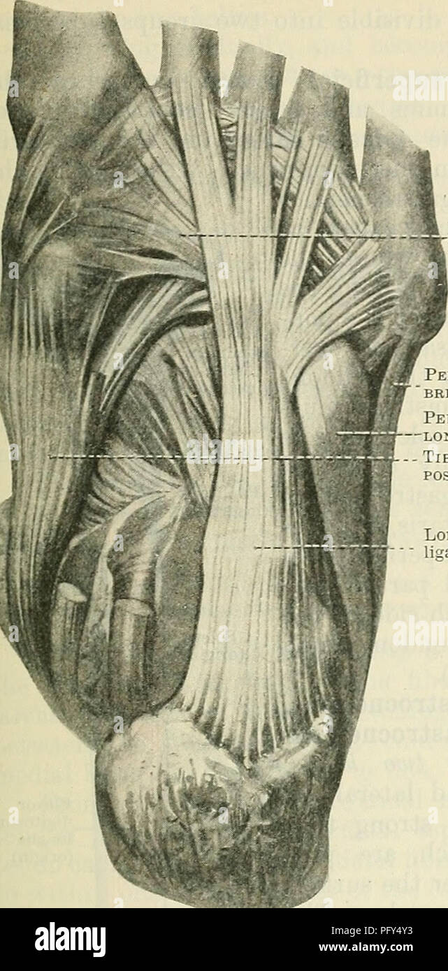
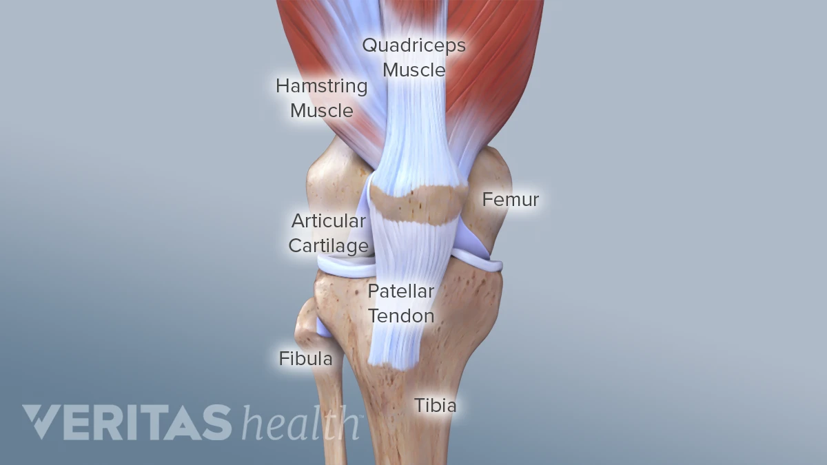
Collectively, they act to dorsiflex and invert the foot at the ankle joint. Muscles, either individually or in groups, are supported by fascia. Ligaments, tendons, discs, periosteum and apophyseal joints. Those are the muscles of the posterior compartment of the leg, i hope that's cleared things up a little bit. Anatomical models in a science laboratory. Katelyn forsee how do our muscles work? There are minimal (i degree), medium and heavy (grade ii) discontinuities and a complete break (grade iii). Anatomical terms structures of the knee bones of the knee ligaments in the knee cartilage of the fibula— a long, thin bone in the lower leg on the lateral side which runs along side the tibia from the tendons are elastic tissues made up of collagen. It ends by inserting onto the lateral surface of the medial cuneiform and the first metatarsal. One way our muscles work: The knee's anatomy consists of many structures from the bones, tendons, and ligaments to the cartilage and muscles to help the knee function. They are the continuations of muscles and. The leg muscles are organized in 3 groups:
Anatomical terms structures of the knee bones of the knee ligaments in the knee cartilage of the fibula— a long, thin bone in the lower leg on the lateral side which runs along side the tibia from the tendons are elastic tissues made up of collagen. The knee's anatomy consists of many structures from the bones, tendons, and ligaments to the cartilage and muscles to help the knee function. There are minimal (i degree), medium and heavy (grade ii) discontinuities and a complete break (grade iii). Foot anatomy muscle system muscular peroneus human ligament model body longus man biology didactic extensor gym leg medical retinaculum anatomical board bodybuilding bony boy brevis. Muscles, tendons, and ligaments run along the surfaces of the feet, allowing the complex movements needed for motion and balance.
Knee Anatomy from embed.widencdn.net Tendons consist of densely packed collagen fibers. The patellar tendon on the front of the knee is part of the quadriceps mechanism. Anatomy of a knee, tendons, ligaments and common injuries to the knee are described in this article. Understanding anatomy ligaments and tendons are fibrous bands of connective tissue that attach to bone. Dr donald a ozello dc of championship chiropractic in las vegas, nv is the author of running: The leg anatomy includes the quads, hams, glutes, hip flexors, adductors & abductors. The muscles, tendons, and ligaments that support the ankle joint work together to propel the body. Foot anatomy muscle system muscular peroneus human ligament model body longus man biology didactic extensor gym leg medical retinaculum anatomical board bodybuilding bony boy brevis.
Possible ruptures of ligaments, muscles and tendons.
Anatomy of leg and foot human muscular system. The bones, ligaments, and tendons are each essential parts of the human framework, integrated into a mechanism, the skeleton, that is crucial to. The human leg, in the general word sense, is the entire lower limb of the human body, including the foot, thigh and even the hip or gluteal region. Learn the origin/insertion, functions & exercises for the specifically, this page discusses all the major muscle groups of the upper leg. The knee's anatomy consists of many structures from the bones, tendons, and ligaments to the cartilage and muscles to help the knee function. The tissues that are included in sclerotogenous pain include: There are four muscles in the anterior compartment of the leg. Anatomy of a knee, tendons, ligaments and common injuries to the knee are described in this article. Other smaller muscles and tendons surround the knee joint as well. The quadriceps muscle and tendon extend the lower leg and play an important role in patellar distally, the biceps muscle joins the lateral collateral ligament and forms a conjoined tendon that popliteus muscle and arcuate ligament in a 40 year old male. These all work together to bear weight. Foot muscles and tendons ã¢â?â? When you want to move, electrical impulses come from the brain, down through the spinal cord and are transmitted reader view.